Thrombophilia
Spontaneous thromboembolic diseases are observed in presence of congenital or acquired deficiencies of antithrombin. These deficiencies are classified in 4 different groups:
• Type I: Decreased AT concentration and decreased AT activity; this is the most frequent case.
• Type II RS (Reactive Site): Normal AT concentration and decreased biological activity; a protein abnormality is present at the active site.
• Type II HBS (Heparin Binding Site): Normal AT concentration, normal AT activity in the absence of heparin, but decreased in its presence.
• Type II (Pleiotropic): Decreased AT concentration and activity; nonfunctional protein and at a lowered level.
* cf Dedicated instructions for use
-
BIOPHEN™ AT anti-(h)-Xa LRTLiquid and Ready to use
Determination of AT activity in human citrated plasma, using an anti-Xa chromogenic method (Human Xa).
Reference Registration Presentation 221123 CE-IVD R1: 3 x 3 mL
R2: 3 x 3 mL221127 CE-IVD R1: 4 x 7.5 mL
R2: 4 x 7.5 mL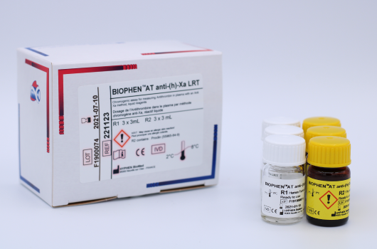
-
BIOPHEN™ AT LRTLiquid and Ready to use
Determination of the heparin cofactor activity of AT in human citrated plasma, using an anti-Xa chromogenic method (Bovine Xa).
Reference Registration Presentation 221111 CE-IVD R1: 4 x 11 mL
R2 : 4 x 4 mL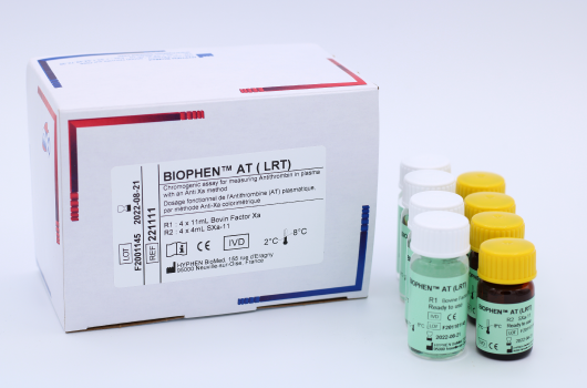
-
BIOPHEN™ Antithrombin
Determination of AT activity in human citrated plasma, using an anti-Xa chromogenic method (Human Xa).
Reference Registration Presentation 221102 CE-IVD
510(k)R1: 2 x 2.5 mL
R2: 2 x 2.5 mL
R3: 2 x 5 mL221105 CE-IVD
510(k)R1: 4 x 5 mL
R2: 4 x 5 mL
R3: 4 x 10 mL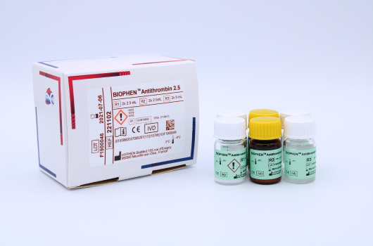
-
BIOPHEN™ AT (Anti-IIa)
Determination of AT activity, in presence of an excess of heparin, in human citrated plasma or purified milieu, using an anti-IIa chromogenic method (Bovine IIa).
Reference Registration Presentation 221122 CE-IVD R1: 2 x 2.5 mL
R2: 2 x 2.5 mL
R3 : 2 x 25 mL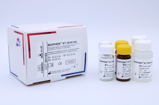
-
LIAPHEN™ AT
 Liquid and Ready to use
Determination of Antithrombin antigen (AT:Ag) in human citrated plasma using a latex turbidimetric assay.
Reference Registration Presentation 120002 CE-IVD R1: 2 x 2 mL
R2: 2 x 10 mL120008 CE-IVD R1: 2 x 1.5 mL
R2: 2 x 5 mL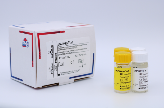
Assay of coagulation PC in plasma helps for the diagnosis of congenital or acquired PC deficiencies.
Acquired deficiencies are observed in hepatic diseases, during VKA therapy or in Disseminated Intravascular Coagulation (DIC).
Congenital deficiencies can be quantitative (Type I) or qualitative (Type II) and are associated with recurrent venous thromboses.
Congenital or acquired PC deficiency is a risk factor for venous thrombosis.
* cf Dedicated instructions for use
-
HEMOCLOT™ Protein C
Determination of PC in human citrated plasma with a clotting method.
Reference Registration Presentation CK031K CE-IVD R1: 3 x 1 mL
R2: 3 x 1 mLCK032K CE-IVD R1: 3 x 2 mL
R2: 3 x 2 mL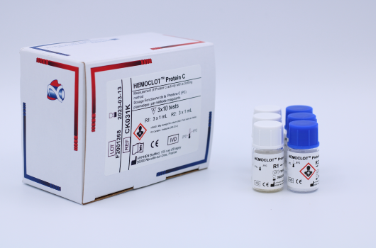
-
BIOPHEN™ Protein C LRTLiquid and Ready to use
Determination of PC activity in human citrated plasma with a chromogenic assay, in liquid format.
Reference Registration Presentation 221211 CE-IVD R1: 3 x 3 mL
R2: 3 x 3 mL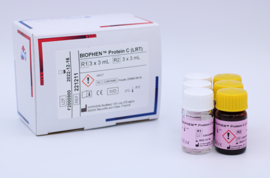
-
BIOPHEN™ Protein C 5
Determination of PC activity in human citrated plasma with a chromogenic assay.
Reference Registration Presentation 221205 CE-IVD
510(k)R1: 4 x 5 mL
R2: 4 x 5 mL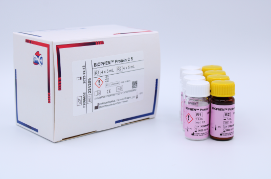
-
ZYMUTEST™ Protein C
Determination of human PC antigen in human plasma by Elisa.
Reference Registration Presentation RK027A CE-IVD 96 tests 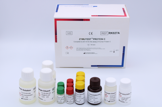
-
Protein C Deficient Plasma
Lyophilized, human citrated plasma, deficient for PC, for any protocol or research study where a source of human PC deficient plasma is required.
Reference Registration Presentation DP100K RUO 6 x 1 mL 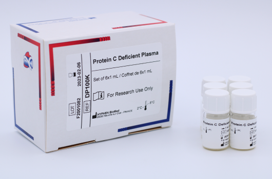
-
LIAPHEN™ Protein CDetermination of Protein C antigen in human citrated plasma.
Reference Registration Presentation 120003 CE-IVD R1: 4 x 5 mL
R2: 4 x 2.3 mL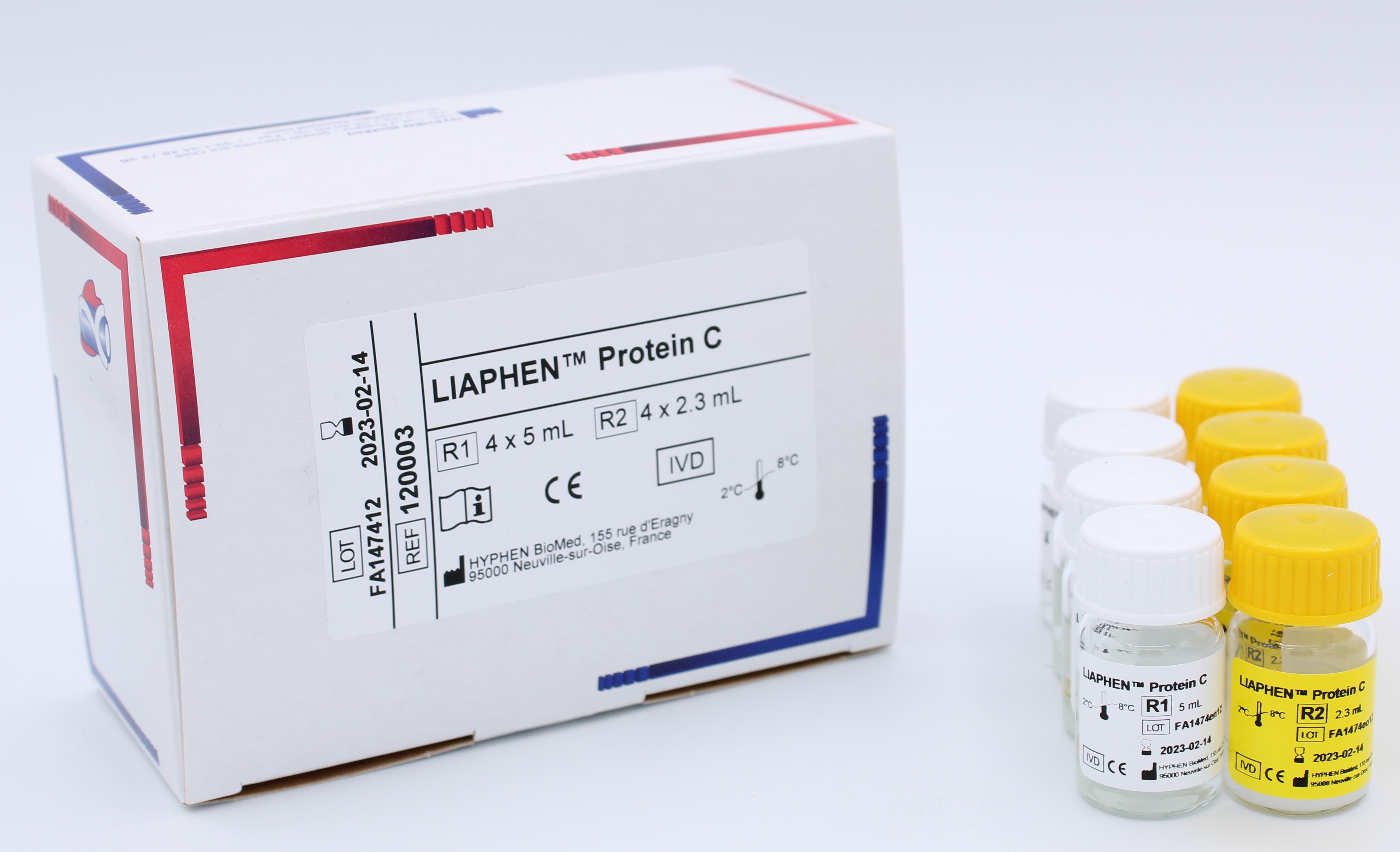
Congenital or acquired PS deficiencies are associated with an increased risk of venous thromboembolism.
In the early stages of inflammatory diseases, free PS concentration is decreased because of an elevation of C4b-BP. PS may be decreased in various contexts such as dicoumarol or L-asparaginase therapy, in hepatic disorders, nephrotic syndrome, during pregnancy, related to oral contraceptives intake or œstrogen therapy, viral infections, DIC. Free PS may also be decreased in new-borns. PS is slightly lower in females than in males.
Inherited PS deficiencies are classified into three types:
• Type I: decreased total and free PS levels (quantitative defects).
• Type II: decreased PS activity but normal antigenic levels (rare qualitative defects).
• Type III: normal levels of total PS but decreased free PS (free form distribution defect).
* cf Dedicated instructions for use
-
LIAPHEN™ Free Protein SLiquid and Ready to use
Determination of free PS Antigen (free PS:Ag) in human citrated plasma with a latex turbidimetric assay.
Reference Registration Presentation 120004 CE-IVD R1: 4 x 3 mL
R2: 4 x 4.4 mL
-
HEMOCLOT™ Protein S
Determination of PS activity in citrated human plasma with a clotting method.
Reference Registration Presentation CK041K CE-IVD R1: 3 x 1 mL
R2: 3 x 1 mLCK042K CE-IVD R1: 3 x 2 mL
R2: 3 x 2 mL
-
ZYMUTEST™ Free Protein S (II)
Determination of Free PS (the activated Protein C (aPC) cofactor) in human plasma by Elisa.
Reference Registration Presentation RK015B
on-demandCE-IVD 96 tests -
ZYMUTEST™ Total Protein S
Measurement of human Total PS in plasma, by Elisa.
Reference Registration Presentation RK021A CE-IVD 96 tests 
-
Protein S Deficient Plasma
Lyophilized, human citrated plasma, deficient for PS, for any protocol or research study where a source of human PS deficient plasma is required.
Reference Registration Presentation DP110K RUO 6 x 1 mL 
Factor V Leiden (FV-L) mutation is the most common hereditary thrombophilia risk factor. Patients carrying FV-L mutation have an increased risk of venous thrombosis.
This genetic anomaly can be evidenced with clotting assays, performed in presence or absence of APC.
* cf Dedicated instructions for use
-
HEMOCLOT™ Quanti. VL
Quantitative determination of FV-L, by measuring its resistance to the inactivation by APC, in the presence of PS, in human citrated plasma.
Reference Registration Presentation CK065K CE-IVD
510(k)R1: 3 x 2 mL
R2: 3 x 1 mL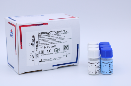
-
ZYMUTEST™ Protein Z
Measurement of human PZ in plasma, by Elisa.
Reference Registration Presentation RK031A RUO 96 tests 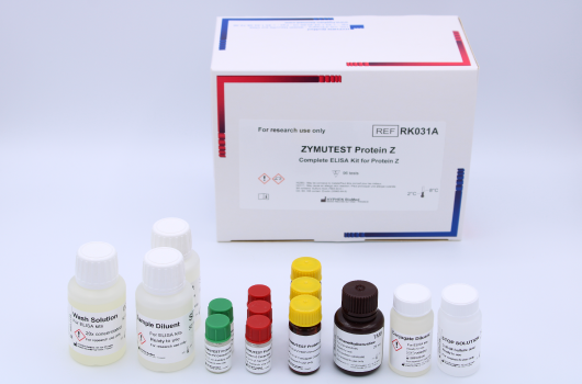
LA are associated with numerous clinical states including e.g. lupus, autoimmune diseases, thrombosis, fœtal loss and must usually be confirmed from multiple assays.
* cf Dedicated instructions for use
-
CEPHEN™Liquid aPTT Ready to use
Activated Partial Thromboplastin Time (aPTT) with low sensitivity to the presence of LA.
Reference Registration Presentation CK511K CE-IVD 6 x 1 mL CK512K CE-IVD 6 x 2.5 mL CK515K CE-IVD 8 x 5 mL CK515L CE-IVD 12 x 5 mL 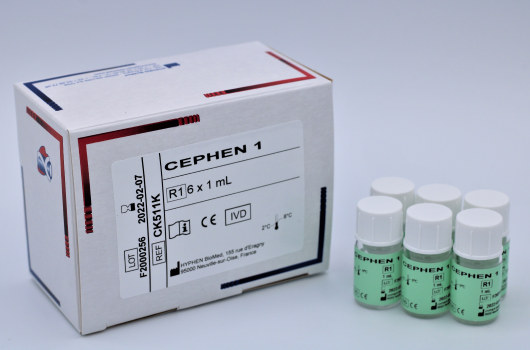
-
CEPHEN™ LSLiquid aPTT Lupus sensitive Ready to use
Activated Partial Thromboplastin Time (aPTT) with high LA sensitivity, to confirm its presence.
Reference Registration Presentation CK521K CE-IVD 6 x 1 mL CK522K CE-IVD 6 x 2.5 mL 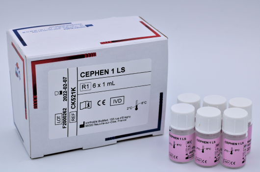
-
HEMOCLOT™ LA-S
Simplified diluted Russell’s Viper Venom Test (dRVVT) to screen for the presence of LA.
Reference Registration Presentation CK090K CE-IVD 6 x 1 mL CK094K CE-IVD 12 x 2 mL 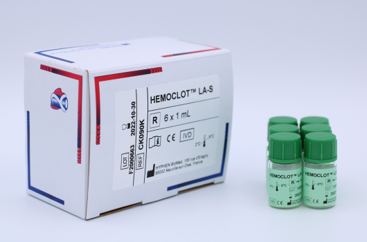
-
HEMOCLOT™ LA-C
Simplified diluted Russell’s Viper Venom Test (dRVVT) with high Phospholipid content to confirm for the presence of LA.
Reference Registration Presentation CK091K CE-IVD 6 x 1 mL 
-
BIOPHEN™ NormalPlasma
Pool of normal human plasma.
Reference Registration Presentation 223602 CE-IVD 10 x 2 mL 223605 CE-IVD 8 x 5 mL 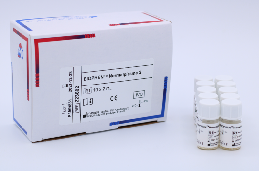
-
ZYMUTEST™ ACA-APA - IgG - Isotype
Measurement of anti-cardiolipin / anti-phospholipid antibodies of the IgG isotype, in human plasma or serum, or in any biological fluid.
Reference Registration Presentation RK029A
on-demandCE-IVD 96 tests 
-
ZYMUTEST™ ANTI β2-GLYCOPROTEIN I - IgG – Isotype
Measurement of autoantibodies to β2GPI of the IgG isotype, by Elisa.
Reference Registration Presentation RK014A
on-demandCE-IVD 96 tests 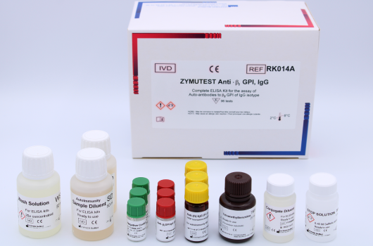
-
ZYMUTEST™ Anti-Annexin V, IgG
Measurement of autoantibodies to Annexin V of the IgG isotype, in human plasma.
Reference Registration Presentation RK005A
on-demandRUO 96 tests 
-
ZYMUTEST™ Anti-Annexin V, IgM
Measurement of autoantibodies to Annexin V of the IgM isotype, in human plasma.
Reference Registration Presentation RK005B
on-demandRUO 96 tests 
-
ZYMUTEST™ Anti–PROTEIN S IgG–Isotype
Measurement of autoantibodies to PS of the IgG isotype, in human plasma.
Reference Registration Presentation RK020A
on-demandRUO 96 tests 
-
ZYMUTEST™ Anti–PROTEIN S IgM–Isotype
Measurement of autoantibodies to PS of the IgM isotype, in human plasma.
Reference Registration Presentation RK020B
on-demandRUO 96 tests 
-
ZYMUTEST™ Anti–PROTEIN Z IgG–Isotype
Measurement of autoantibodies to Protein Z of the IgG isotype, in human plasma.
Reference Registration Presentation RK025A
on-demandRUO 96 tests 
-
ZYMUTEST™ Anti–PROTEIN Z IgM–Isotype
Measurement of autoantibodies to Protein Z of the IgM isotype, in human plasma.
Reference Registration Presentation RK025B
on-demandRUO 96 tests 
-
ZYMUTEST™ Anti-Prothrombin IgG
Measurement of autoantibodies to Prothrombin, of the IgG isotype, in human plasma.
Reference Registration Presentation RK007A
on-demandRUO 96 tests 
-
ZYMUTEST™ β2GPI
Immuno-assay for measuring human Beta 2 Glyocoprotein I (β2GPI) in plasma, by Elisa.
Reference Registration Presentation RK022A
on-demandRUO 96 tests 
-
ZYMUTEST™ Annexin V
Measurement of human Annexin V in plasma, by Elisa.
Reference Registration Presentation RK004A
on-demandRUO 96 tests 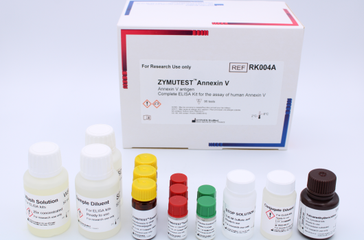
-
ZYMUTEST™ DDimer
Measurement of the Fibrin Degradation Products (DDimer) in plasma, by Elisa.
Reference Registration Presentation RK023A
on-demandRUO 96 tests 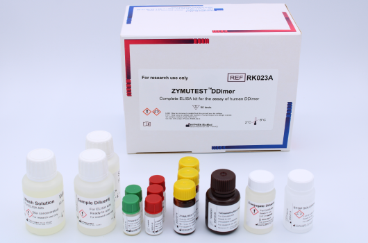
-
ZYMUTEST™ FPA
Determination of human FPA, for use on bentonite adsorbed human plasma, using a Competitive Enzyme Linked Immuno Assay (CELIA). Fibrinogen, when present in the specimen, must be removed (i.e., bentonite adsorption of human plasma), as it cross-reacts with antibodies to FPA.
Reference Registration Presentation RK016B RUO 96 tests 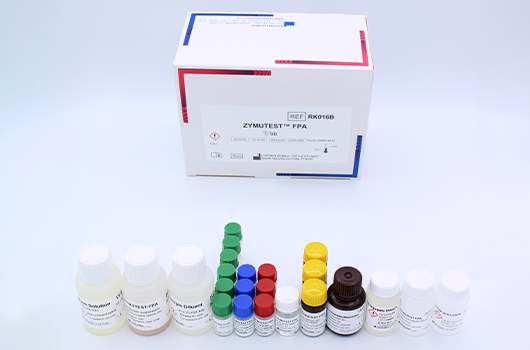
-
ZYMUTEST™ CRP
High sensitive measurement of human CRP in plasma, serum, by Elisa.
Reference Registration Presentation RK010A
on-demandRUO 96 tests 
-
ZYMUPHEN™ MP ACTIVITY
Measurement of microparticles’ procoagulant activity in human plasma.
Reference Registration Presentation 521096
on-demandRUO 96 tests 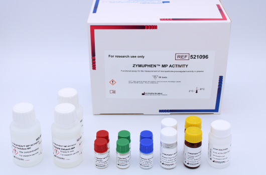
-
ZYMUPHEN™ MP-TF
Determination of the procoagulant activity of microparticles exposing Tissue Factor (MP-TF), in human plasma and in purified milieu.
Reference Registration Presentation 521196
on-demandRUO 96 tests -
ZYMUTEST™ Tissue Factor (full length)
Determination of Full-Length Tissue Factor (FL-TF) (1-263) on plasma and purified milieu.
Reference Registration Presentation RK035A
on-demandRUO 96 tests 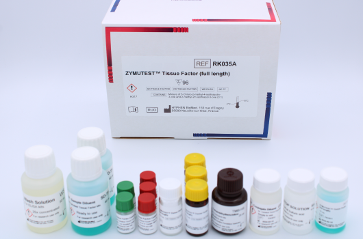
-
ZYMUTEST™ Total Tissue Factor
Determination of Tissue Factor (TF) on plasma and purified milieu. This kit does not recognize alternatively spliced Tissue Factor (asTF).
Reference Registration Presentation RK042A
on-demandRUO 96 tests 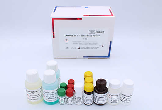
-
BIOPHEN™ Factor XIa
Measurement of activated Factor XI (FXIa) activity, through FIX activation and Factor Xa generation.
Reference Registration Presentation 220412 RUO R1A: 2 x 3 mL
R1B: 2 x 3 mL
R2: 2 x 3 mL
R3: 2 x 3 mL
R4: 2 x 25 mL
Cal: 2 x 2 mL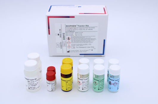
-
BIOPHEN™ Factor IXa
Determination of FIXa activity, in purified medium.
Reference Registration Presentation 221812 RUO R1: 2 x 2.5 mL
R2: 2 x 2.5 mL
R3: 2 x 2.5 mL
R4: 2 x 25 mL
Cal: 2 x 1 mL
-
HEMOCLOT™ Factor VIIa
Determination of activated Factor VII (FVIIa) activity, in purified medium or citrated plasma.
Reference Registration Presentation CK092K CE-IVD R1: 3 x 2 mL
R2: 3 x 2 mL
R3: 3 x 20 mL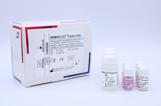
-
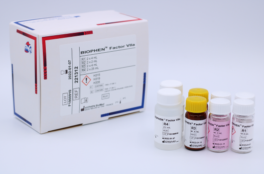
BIOPHEN™ Factor VIIa
Determination of activated Factor VII (FVIIa) activity, in purified milieu.
Reference Registration Presentation 221312
on-demandRUO R1: 2 x 4 mL
R2 : 2 x 2 mL
R3 : 2 x 4 mL
R4 : 2 x 25 mL